







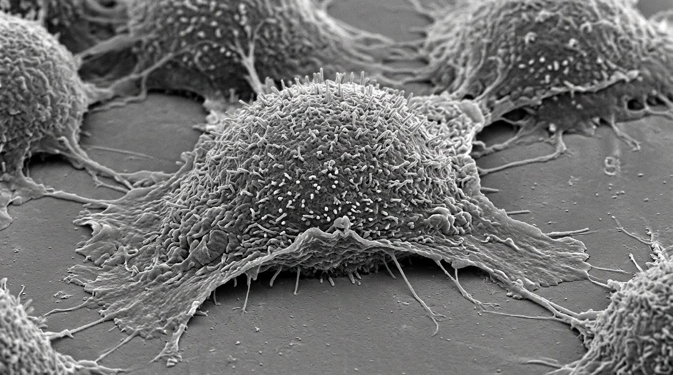
A laboratory investigation has demonstrated that gold nanoparticles synthesized using a secondary metabolite isolated from the lichen Parmotrema reticulatum exhibit targeted cytotoxic activity against cultured human lung cancer cells. The study, documented by an academic research team, focused on fabricating novel nanoformulations through a green nanotechnology route that impregnates gold nanoparticles with a specific dioxepine derivative. In vitro experiments conducted on the human lung cancer cell line A549 revealed that the resulting surface-capped nanoparticles induced programmed cell death, triggered nuclear condensation and fragmentation, and arrested the cellular division cycle during the synthesis phase, yielding a half-maximal inhibitory concentration of 12 micrograms per milliliter.
The biological material serving as the capping agent was extracted directly from specimens of Parmotrema reticulatum, a lichen species belonging to the Parmeliaceae family that naturally colonizes trees and rock surfaces and occurs prominently in the Eastern Ghats of India. Researchers recovered the active chemical constituent through Soxhlet extraction followed by column chromatography over silica gel using varied proportions of polar and nonpolar solvents, verifying fractions via thin-layer chromatography. Comprehensive spectral characterization employing Fourier-transform infrared spectroscopy, high-performance liquid chromatography on a C18 column, proton and carbon nuclear magnetic resonance spectroscopy, two-dimensional correlation spectroscopy, and liquid chromatography-mass spectrometry confirmed the isolated molecule as 3-hydroxy-9-hydroxymethyl-1,8-dimethyl-11-oxo-11H-dibenzo[b,e][1,4]dioxepine-4,6-dicarboxylic acid 6-methyl ester, having the molecular formula C19H16O9 and an identified molecular weight of 388.34 grams per mole.
To synthesize the hybrid nanostructures, the investigators dissolved the purified dioxepine derivative in distilled water at a concentration of 1 milligram per milliliter and introduced auric chloride trihydrate at a final precursor concentration of 2 millimolar. The reaction mixture was agitated overnight on a magnetic stirrer, during which an overt optical shift from green to cherry red confirmed the reduction and formation of dioxepine-derived gold nanoparticles. Following incubation, the colloidal suspension was subjected to centrifugation, and the collected nanoparticle pellet underwent multiple washing stages with distilled water to thoroughly eliminate unreacted chemical reagents, residual precursors, and soluble reaction by-products prior to subsequent analytical testing.
Initial optical characterization using ultraviolet-visible spectrophotometry established a pronounced surface plasmon resonance peak centered at 572 nanometers, corroborating complete colloidal dispersion of the synthesized gold nanoparticles within the aqueous medium. Vibrational analysis using Fourier-transform infrared spectroscopy identified prominent absorption bands that clarified the organic interface capping the metallic cores. A broad band observed at 3461 inverse centimeters confirmed the presence of hydroxyl functional groups, while an absorption feature at 2928 inverse centimeters corresponded to the stretching vibrations of carbohydrate aliphatic carbon-hydrogen bonds. A sharp spectral peak at 1660 inverse centimeters was assigned to carbonyl stretching vibrations within ester linkages, accompanied by specific bands at 1389, 1262, and 1181 inverse centimeters attributable to carbon-oxygen stretching modes of alcohols and esters, reflecting the coordination of hydroxyl and carbonyl groups during gold stabilization.
Crystallographic examination using X-ray diffraction resolved three distinct diffraction peaks positioned at 2-theta angles of 38.55 degrees, 46.63 degrees, and 64.80 degrees. These reflections correspond respectively to the standard Bragg planes of (111), (200), and (220) belonging to the face-centered cubic lattice geometry of zero-valent gold indexed under standard powder diffraction reference JCPDS 65-2870. The dominant reflection intensity documented at 38.55 degrees evidenced preferential crystalline growth oriented along the (111) direction. Application of the Scherrer equation to the experimental diffraction profile determined that the engineered nanoparticles possessed an average crystallite domain size of 26.8 nanometers, with minor accessory peaks reflecting surface-bound bioorganic capping constituents.
Microscopic analyses provided detailed dimensional and morphological profiles of the synthesized formulation. Scanning electron microscopy resolved predominantly spherical particles dispersed throughout the solution with dimensions measuring between 25 and 30 nanometers. High-resolution transmission electron microscopy expanded these observations, capturing a mixture of spherical, triangular, and hexagonal geometric profiles with individual particle sizes spanning from 20 to 30 nanometers. Selected area electron diffraction patterns generated during transmission imaging displayed characteristic concentric polycrystalline rings that substantiated the polycrystalline nature of the quasi-spherical and triangular metallic architectures.
Hydrodynamic profiling of the colloidal suspension via dynamic light scattering recorded an average hydrodynamic diameter of 661 plus or minus 0.8 nanometers, exhibiting a predominant peak intensity of 88 percent. The measured polydispersity index reached 0.625, illustrating a polydisperse distribution attributable to structural shape heterogeneity and the presence of hydration spheres in aqueous suspension. Complementary surface electrokinetics determined through zeta potential measurements yielded a positive value of plus 58 millivolts. This pronounced positive surface charge provides electrostatic repulsion that impedes particle agglomeration in suspension, indicating colloidal stability mediated by the organic surfactant layer surrounding the gold metallic core.
The radical-scavenging properties of the nanoformulation were quantified using two independent in vitro antioxidant procedures across a concentration gradient from 20 to 100 micrograms per milliliter. In the 2,2-diphenyl-1-picrylhydrazyl assay, the nanoparticles demonstrated dose-dependent radical quenching ranging from 21 percent to 45 percent, culminating in 63.68 percent inhibition at peak experimental concentrations as electron transfer reduced the radical from violet to yellow. Parallel testing via the ferric ion reducing antioxidant power assay measured an increase in reduction capacity from 25.7 percent to 40.96 percent across the identical concentration range, registering an overall assay response between 12 percent and 71 percent. The authors noted that this ferric-reducing capacity was lower than that of the uncomplexed dioxepine molecule, pointing toward an antagonistic interaction during the reduction of ferric iron to ferrous iron within that specific assay format.
Evaluation of biological activity against human non-small cell lung cancer cells was performed using the A549 line maintained in Dulbecco’s modified Eagle’s medium supplemented with fetal bovine serum and antibiotics. Cytotoxicity measured through colorimetric 3-(4,5-dimethylthiazol-2-yl)-2,5-diphenyltetrazolium bromide assays established that nanoparticle exposure caused concentration-dependent reductions in cell viability, yielding an IC50 benchmark of 12 micrograms per milliliter alongside comparative doxorubicin controls. Bright-field inverted microscopy documented progressive, dose-dependent cytoskeletal deterioration across concentrations of 20, 40, and 80 micrograms per milliliter, manifesting visible hallmarks of cell death including cellular shrinkage, shape deformation, cell clumping, and membrane blebbing compared to untreated control monolayers.
Fluorescence microscopy elucidated the mode of cytotoxicity induced by the lichen-capped gold nanoparticles. Acridine orange and ethidium bromide dual staining revealed viable cells with intact membrane integrity emitting uniform green fluorescence, whereas treated cancer cells displayed yellow-green emission indicative of early apoptosis alongside orange-to-red fluorescence marking late-stage apoptotic and necrotic cells compromised by loss of membrane integrity. Complementary nuclear staining utilizing 4′,6-diamidino-2-phenylindole exposed widespread chromatin condensation and nuclear DNA fragmentation that intensified in direct proportion to the nanoparticle dosage administered to the culture wells.
To establish the exact phase of growth inhibition, the researchers conducted cell cycle distribution analyses using propidium iodide staining and flow cytometry on treated A549 populations. The resulting DNA content profiles indicated that exposure to the nanoparticles selectively hindered cell cycle progression by inducing an arrest specifically within the synthesis phase, thereby arresting DNA replication. Subsequent protein quantification by Western blot confirmed that this cell cycle blockade coincided with the molecular execution of the intrinsic apoptotic pathway, marked by the activation and cleavage of caspase-9, an initiator regulatory enzyme linked to apoptosome formation and mitochondrial membrane rupture.
While these laboratory findings show that dioxepine-conjugated gold nanoparticles exert potent antiproliferative, apoptotic, and antioxidant effects in cultured lung cancer cells, the authors explicitly contextualized the results as preliminary in vitro findings. The study evaluated isolated cellular lines under controlled bench conditions and did not assess pharmacokinetic behavior, systemic toxicity, or therapeutic efficacy within living organismal models. Consequently, the research team emphasized that substantial additional investigation remains necessary before these lichen-functionalized gold nanomaterials can be considered for potential clinical or therapeutic oncology applications.
Subject of Research: Green synthesis and in vitro evaluation of dioxepine-functionalized gold nanoparticles against human lung cancer cells
Article Title: Dioxepine-derived surface-capping gold nanoparticles (Dd-AuNPs) induces ROS-mediated apoptosis and cell cycle arrest in A549 human lung cancer cell line
Article References: Khader, S. Z. A., Syed Zameer Ahmed, S., Raju, M., Mahboob, M. R., Subramaniyan, S., Kaliyannan Rajavel, A., Chinnaperumal, K., & Sakthivel, D. (2024). Dioxepine-derived surface-capping gold nanoparticles (Dd-AuNPs) induces ROS-mediated apoptosis and cell cycle arrest in A549 human lung cancer cell line. Gold Bulletin, 57(1-2), 65-77. https://doi.org/10.1007/s13404-024-00348-4
Image Credits: AI Generated
DOI: 10.1007/s13404-024-00348-4
Keywords: Gold nanoparticles, Parmotrema reticulatum, Dioxepine derivative, A549 cell line, Lung cancer, Green nanotechnology, MTT assay, Apoptosis, Cell cycle arrest, S phase, Caspase-9, Reactive oxygen species, DPPH assay, FRAP assay, Zeta potential, High-resolution TEM, X-ray diffraction, Flow cytometry