







Dynamic alterations in retinal vascular geometry triggered by changes in body posture provide significant prognostic indications for the progression of diabetic retinopathy in individuals with type 2 diabetes mellitus, according to prospective pilot research published in Eye and Vision. Investigators demonstrated that measuring shifts in retinal vessel parameters as participants move from a sitting to a supine position can expose subclinical microvascular dysregulation before advanced structural lesions appear. By implementing a portable smartphone-based retinal imaging adapter alongside semi-automated vascular analysis software, the researchers captured postural vascular responses that independently predicted clinical disease worsening over an average follow-up period of five years. These physiological observations suggest that evaluating dynamic autoregulatory capacity, rather than relying exclusively on conventional static fundus appearance or systemic glycemic metrics, could significantly refine individual risk stratification.
Diabetic retinopathy remains a principal cause of preventable vision impairment and blindness worldwide, operating as a frequent microvascular complication of chronic diabetes mellitus. However, traditional systemic risk factors, including duration of diabetes, blood pressure, and glycated hemoglobin levels, do not fully account for individual variability in disease trajectory. Consequently, standard annual screening protocols can prove unnecessarily frequent for stable individuals while failing to identify those at elevated risk of rapid visual deterioration. A pressing clinical necessity exists for non-invasive functional biomarkers capable of distinguishing latent microvascular vulnerability, thereby permitting timely ophthalmic referral and optimized monitoring schedules in resource-conscious healthcare settings.
The human retina provides an accessible anatomical window where microvascular networks can be directly visualized and measured without invasive interventions. Under healthy physiological conditions, retinal blood flow is carefully stabilized against postural shifts in hydrostatic and perfusion pressures through an intrinsic autoregulatory myogenic response. When an individual transitions from an upright sitting posture to a horizontal supine posture, ocular perfusion pressure rises, prompting normal retinal arterioles to contract compensatorily. In individuals affected by diabetes mellitus, persistent hyperglycemia and subsequent endothelial dysfunction impair this adaptive vasoconstriction, attenuating local autoregulatory capacity. Prior research indicated that a defective myogenic response manifests early in type 1 diabetes, but examining posture-induced microvascular shifts in type 2 diabetes has historically been hindered by the physical bulk and rigidity of standard tabletop fundus cameras, which cannot easily accommodate patients lying flat.
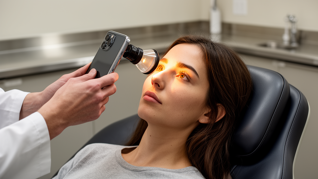
To overcome these physical constraints, researchers utilized a lightweight smartphone setup equipped with a specialized clip-on adapter lens known as the OphthoLens, manufactured by RainsOptics Ltd. This portable optical assembly, positioned directly over the mobile device camera and light source, allowed standardized high-resolution retinal photography across distinct postures. The clinical protocol evaluated participants consecutively enrolled from the Chinese University of Hong Kong Eye Center between August 2018 and August 2019. Eligible subjects included adult patients with confirmed type 2 diabetes mellitus presenting with no retinopathy or non-proliferative diabetic retinopathy, alongside an age-matched cohort of healthy control participants. Individuals with proliferative disease, macular edema, recent intraocular surgery, or secondary ocular pathologies were systematically excluded to isolate baseline microvascular behavior.
The experimental procedure enforced strict positional stabilization to avoid confounding acute hemodynamic fluctuations during data collection. Participants were rested in an upright seated position for ten minutes before image acquisition, followed by an identical ten-minute rest period upon shifting to the supine position on an examination bed. Ophthalmic technicians captured fundus images before and after pharmacological pupil dilation under supervised guidance. Concurrently, systemic blood pressure was documented using a digital brachial monitor, and intraocular pressure was recorded with a portable tonometer in both postures, allowing the precise computation of mean arterial blood pressure and mean ocular perfusion pressure across postural states. A subset of thirty participants underwent repeat imaging to confirm intra-observer reliability and measurement reproducibility.
Quantitative geometric processing of the smartphone fundus photographs was executed using the Singapore I Vessel Assessment software platform, commonly designated as SIVA. This semi-automated system locates the optic disc, applies a standardized polar measurement grid extending between 0.5 and 2.0 disc diameters from the disc margin, and delineates arterioles from venules. Graders verified automated vessel tracings, quantifying parameters that included vessel caliber, fractal dimension, vessel tortuosity, and branching architecture. Cross-sectional comparisons between thirty-eight healthy controls and forty-nine participants with diabetes examined baseline response profiles using paired and independent testing. In longitudinal evaluations, participants with diabetes were followed prospectively over a median duration of 59.5 months to identify diabetic retinopathy progression, defined by a two-step or greater advancement on the Early Treatment Diabetic Retinopathy Study severity scale.
Cross-sectional evaluations demonstrated striking physiological disparities in positional vascular responses between healthy eyes and diabetic eyes. In healthy controls, transitioning from the sitting to the supine posture elicited standard physiological constriction in both retinal arterioles, which narrowed from 92.48 micrometers to 89.32 micrometers, and venules, which decreased from 130.19 micrometers to 124.89 micrometers. Conversely, participants presenting with diabetes and baseline retinopathy exhibited no statistically significant caliber reduction, displaying mean arteriolar calibers of 111.55 micrometers sitting versus 114.69 micrometers supine, and venular calibers of 160.84 micrometers sitting versus 161.89 micrometers supine. Adjusted percentage analyses demonstrated a statistically significant linear trend across controls, diabetic patients without retinopathy, and diabetic patients with retinopathy, tracking a shift from healthy arteriolar constriction of minus 2.10 percent to paradoxical vasodilation reaching plus 6.46 percent.
During the five-year longitudinal observation window, fourteen eyes belonging to the diabetes cohort experienced confirmed disease progression. Proportional hazards modeling identified specific baseline postural adaptations as independent prognostic determinants of retinopathy progression after adjusting for baseline age, glycated hemoglobin, mean arterial pressure, diabetes duration, and baseline retinopathy grade. Specifically, an amplified increase in retinal arteriolar tortuosity during the sitting-to-supine transition correlated with a 2.41-fold elevated risk of retinopathy worsening, yielding a multivariable hazard ratio of 2.41 with a 95 percent confidence interval spanning 1.37 to 4.23. Conversely, wider postural retinal venular branching angles conferred a protective association, reflecting an estimated 45 percent reduction in progression risk with a hazard ratio of 0.55 and a 95 percent confidence interval from 0.35 to 0.87.
Evaluating discriminatory performance indicated that incorporating posture-derived microvascular parameters substantively improved prognostic accuracy compared to risk models constructed solely from traditional clinical variables. Adding baseline percentage adjustments in arteriolar tortuosity increased the model concordance statistic from 0.630 to 0.740, representing a statistically significant enhancement confirmed by likelihood ratio testing. Similarly, incorporating postural venular branching angle changes raised the C-statistic to 0.683. The exaggerated tortuosity observed in vulnerable microvessels likely mirrors severe endothelial dysfunction and reduced nitric oxide bioavailability, preventing structural stability under altered perfusion pressures, whereas flexible venular branching angles may reflect preserved architectural compliance capable of dissipating fluid mechanical stresses.
While the study demonstrates the feasibility of combining smartphone-based optics with computational vascular geometry, several methodological limitations characterize the findings. The investigation operated as an exploratory single-center pilot involving a modest sample of forty-nine diabetic individuals, meaning that the observed associations warrant confirmation in larger multicenter cohorts. Additionally, the limited number of progression events precluded comprehensive subgroup analyses concerning incident retinopathy development among patients entirely free of baseline lesions. The authors also noted that detailed records concerning specific vasoactive glucose-lowering agents, such as sodium-glucose cotransporter-2 inhibitors or glucagon-like peptide-1 receptor agonists, were unavailable. Nonetheless, this dynamic mobile imaging approach illustrates how portable hardware could ultimately support risk stratification and telemedicine screening across diverse community settings.
Subject of Research: Smartphone-based retinal vascular imaging and diabetic retinopathy progression
Article Title: Postural changes in retinal vascular parameters and risk of diabetic retinopathy progression in type 2 diabetes mellitus: a pilot study
Article References: Nguyen, T. X., Wang, Y. M., Ling, A., Zheng, C., Lai, K. H. W., Tang, Z., Liu, J. H. K., & Cheung, C. Y. (2026). Postural changes in retinal vascular parameters and risk of diabetic retinopathy progression in type 2 diabetes mellitus: a pilot study. Eye and Vision, 13(1), Article 3. https://doi.org/10.1186/s40662-025-00471-z
Image Credits: AI Generated
DOI: 10.1186/s40662-025-00471-z
Keywords: Diabetic Retinopathy, Type 2 Diabetes, Smartphone Fundus Imaging, Retinal Microvasculature, Postural Hemodynamics, Arteriolar Tortuosity, Venular Branching Angle, Ocular Autoregulation, Microvascular Complications, SIVA Software, Endothelial Dysfunction, Clinical Ophthalmology, Predictive Biomarkers, Myogenic Response, Fundus Photography, Disease Progression